Photophysical mechanisms and methodological developments P. Didier & Y. Mély

This research axis is focused on the detailed characterization of the photophysics that govern the emission of i) fluorescent nucleoside analogues and ii) oxyluciferin; iii) the properties of upconverting nanoparticles and iv) the application of various methodological developments in relation to quantitative and high resolution microscopy techniques.

1. Photophysics of thienoguanosine, an outstanding fluorescent analogue of guanosine

Interactions of nucleic acids with DNA- and RNA-binding proteins play a key role in transcription, translation, regulation of RNA stability and turnover, and many other cellular processes. A large number of structures of nucleic acid–protein complexes have become available mainly by X-ray crystallography and cryoelectron microscopy, but these methods mostly provide static snapshots of the highly dynamic complexes. Complementary techniques are thus required to study the binding in real time and to probe the dynamics of the complexes. Fluorescence-based methods are highly suited for studying the dynamics and interactions of biological molecules in solution. Unfortunately, the natural nucleobases in nucleic acids are virtually non-fluorescent under ambient conditions, so that extrinsic fluorescent reporters are needed. Multiple strategies have been proposed to fluorescently label nucleic acids. So far, the most suited but also the most chemically demanding approach consists in position-specific replacement of natural nucleobases by fluorescent nucleoside analogues (FNAs), such as 2-aminopurine. However, most FNAs developed so far either perturb the native structure of the labelled nucleic acid and/or are strongly quenched when incorporated in nucleic acids.

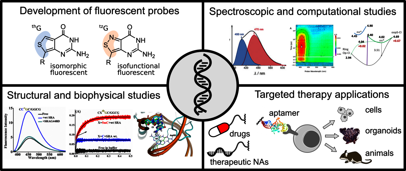
In this context, a breakthrough has been achieved with the introduction by the lab of Y. Tor (UCSD) of the thieno[3,4-d]-pyrimidine family, and notably thienoguanosine (thG). In collaboration with Y. Tor, we made a side by side comparison of thG with 2Ap in its ability to substitute a natural guanosine residue in the (-)DNA copy of the HIV-1 primer binding site, (-)PBS, both in its stem loop conformation and in the corresponding (-)/(+)PBS duplex (1). In contrast to 2Ap, when included in (-)PBS or (-)/(+)PBS duplex, thG fully preserves their stability and exhibits a respectable quantum yield (QY) and a simple fluorescence decay, with no dark species. In further contrast to 2Ap, thG reflects the predominantly populated G conformers, which allows exploring their relevant dynamics. Thus, thG selectively and faithfully monitors the conformations and dynamics of a given G residue in a DNA sequence. In collaboration with Y. Tor, M. Mori (U. Siena) and R. Improta (U. Naples), we also showed that as a free probe, thG exists in two tautomeric forms, identified as the H1, being the only one observed in nonprotic solvents, and H3 keto-amino tautomers (2). These two tautomers exhibit similar QYs, but shifted absorption and emission spectra, as well as different fluorescent lifetimes. Both tautomers are observed when thG is incorporated in single-stranded sequences, while the H1 tautomer is strongly favored in DNA duplexes. These tautomers and their distinct environmental sensitivity provide unprecedented information channels for analyzing G residues in oligonucleotides and their complexes. For both tautomers, the minimum of the spectroscopic state is separated from the most easily accessible conical intersection by a sizeable energy barrier (≥0.4 eV), which explains their large fluorescence QY (3). We further explored the photophysical properties of thG as a function of pH4. This enabled us to identify three additional thG forms, resulting from pH-dependent ground-state and excited-state reactions, and to propose a complete photoluminescence pathway of thG in aqueous solution.

Finally, we thoroughly investigated a series of DNA duplexes, where the bases facing and neighboring thG were systematically varied, to obtain a full understanding of its photophysics in DNA. In matched duplexes, thG's fluorescence QY and lifetime values were almost independent of the flanking bases. The high duplex stability likely maintains a steady orientation and distance between nucleobases, so that a similar charge transfer (CT) mechanism governs thG’s photophysics independently of its flanking nucleobases. In contrast, the local destabilization induced by a mismatch or an abasic site restores a strong dependence of thG's QY and lifetime values on its environmental context, depending on the CT route efficiency and solvent exposure of thG. Thus, thG is perfect for monitoring local structural changes and single nucleotide polymorphism. Moreover, thG's dominant fluorescence lifetime in DNA is unusually long (9-29 ns), facilitating its selective measurement using a lifetime-based or a time-gated detection scheme. Thus, thG is an outstanding emissive substitute for G with good QY, long fluorescence lifetimes, and exquisite sensitivity to local structural changes (5).

Based on its unique properties, thG was used to monitor the annealing mechanism of (+)/(-)PBS6, the NCp7-mediated exposure to solvent of the G7 residue in (-)PBS1 and the UHRF1-mediated base flipping of methylcytosine in hemi-methylated DNA (7).

2. Oxyluciferine

Bioluminescence is a natural phenomenon during which living organisms convert chemical energy into light. The light is emitted through luciferase-catalyzed reaction of D-luciferin substrate resulting in the formation of oxyluciferin (OxyLH2) in its first singlet excited state, which later decays radiatively to the ground state. OxyLH2 emits visible light with an emission that can vary from green to red. It can exist under six different chemical forms resulting from keto/enol tautomerization and deprotonation of phenol and/or enol moieties. We have extensively characterized the optical properties of firefly’s oxyluciferin both experimentally (8–10) and theoretically (11–13). In particular, steady-state and time-resolved spectroscopy were used to disentangle the complex photoluminescence pathways of firefly’s oxyluciferin in solution. We characterized the optical properties of OxyLH2 and its derivatives in aqueous buffer and we deciphered its photoluminescence pathway by monitoring the excited state proton transfer using pump-probe spectroscopy. We are currently characterizing the absorption and emission of OxyLH2’s forms inside the protein.

3. Upconversion nanoparticles

Upconverting nanoparticles (UCNPs) owe their name to the upconversion phenomenon, where high-frequency light is emitted upon sequential absorption of several quanta of low-frequency light. As a result of this Anti-Stokes emission, several critical problems of conventional fluorescent dyes are strongly reduced, namely autofluorescence, excitation light scattering and phototoxicity. Therefore, UCNPs can be imaged with an exceptional signal to noise ratio and used in numerous applications, including low-background imaging, high-sensitivity assays, and cancer theranostics. In these applications, UCNPs are frequently used as a donor in resonance energy transfer (RET) pairs. However, because of the non-linearity of their luminescence mechanism, their behavior as a RET pair component has been difficult to predict quantitatively, preventing their optimization for subsequent applications.

In collaboration with the group of T. Hirsch (U. Regensburg), we assembled UCNP-organic dye RET systems and investigated their luminescence decays and spectra, with varying UCNP sizes and quantities of dyes grafted onto their surface14. We observed an increase in RET efficiency with lower particle sizes and higher dye decoration. We also observed a quenching of UCNP luminescence bands that are not resonant with the absorption of organic dyes. We proposed a semi-empirical Monte Carlo model for predicting the behavior of UCNP-organic dye systems.

Ensemble measurements of diluted aqueous dispersions of UCNPs have shown the instability of luminescence over time due to particle dissolution-related effects. In this context, we have quantified this effect at the individual particle level by using quantitative wide-field microscopy (15). Individual UCNPs exhibit a rapid luminescence loss, accompanied by large changes in spectral response, leading to a considerable heterogeneity in their luminescence and band intensity ratio. This dissolution-related intensity loss is not correlated with the initial particle intensity or band ratio, which makes it virtually unpredictable. These effects can be largely slowed down by adding millimolar concentrations of sodium fluoride in buffer. Thus, microscopy experiments employing UCNPs in aqueous environment should be performed under conditions that carefully prevent these effects.

During the last few years, we have developed and applied a number of methods in quantitative and high resolution microscopy.

4. Methodological developments

4.1 ReAsH/tetracystein-based correlative light-electron microscopy (CLEM) for HIV-1 imaging

Visualization of HIV-1 in infected cells by CLEM requires a specific labelling in order to recognize the nanometric viral cores in the intracellular environment. To reach this aim, we adapted the labeling approach with a tetracystein tag (TC) and a biarsenical resorufin-based label (ReAsH) (16). In this approach, the ReAsH fluorophore triggers the photo-conversion of 3,3-diaminobenzidine tetrahydrochloride (DAB), generating a precipitate sensitive to osmium tetroxide staining that can be visualized by transmission electron microscopy (TEM). The TC tag is fused to the nucleocapsid protein NCp7, that binds to the viral genome. HeLa cells, infected by ReAsH-labeled pseudoviruses containing NCp7-TC proteins exhibit strong fluorescent cytoplasmic spots that overlap with dark precipitates in the TEM sections. Our work opened new perspectives for the use of CLEM to monitor the intracellular traffic of viral complexes.

4.2 Optimized protocol for combined PALM-dSTORM imaging

Multi-colour super-resolution localization microscopy is an efficient technique to study a variety of intracellular processes, including protein-protein interactions. This technique requires specific labels that display transition between fluorescent and non-fluorescent states under given conditions. For the most commonly used label types, namely photoactivatable fluorescent proteins and organic fluorophores, these conditions are different, making experiments that combine both labels difficult. Here, we demonstrate that changing the standard imaging buffer of thiols/oxygen scavenging system, used for organic fluorophores, to the commercial mounting medium Vectashield increased the number of photons emitted by the fluorescent protein mEos2 and enhanced the photoconversion rate between its green and red forms. In addition, the photophysical properties of organic fluorophores remained unaltered with respect to the standard imaging buffer. The use of Vectashield together with our optimized protocol for correction of sample drift and chromatic aberrations enabled us to perform two-colour 3D super-resolution imaging of the nucleolus and resolve its three compartments (17).

4.3 FLIM-FRET use for exploring protein–protein interactions with unbalanced protein expression levels

Förster resonance energy transfer measured by fluorescence lifetime imaging (FLIM-FRET) enables to map protein-protein interactions (PPIs) in a living cell with high spatial and temporal specificity. However, the accurate measurement and interpretation of multi-exponential FLIM-FRET data associated to mixtures of interacting and non-interacting proteins are difficult. We introduced a simple diagram plot that clusters pixels with similar decay signatures (18). FLIM diagram plots can provide valuable information about stoichiometry and binding mode in PPIs, even in the presence of large differences in protein expression levels of the different interacting partners. This FLIM diagram plot is a useful visual approach for straightforward interpretation of complex lifetime data and was successfully applied for revealing protein-protein interactions in live bacteria (19).

4.4 FLIM imaging of membrane lipid order and HIV-1 Gag oligomerization

Lateral segregation of lipids into liquid-ordered (Lo) and -disordered (Ld) phases in lipid membranes can be monitored with environment-sensitive dyes, such as the dual-color F2N12S probe. The two bands of F2N12S are associated with the normal (N*) and tautomer (T*) excited-state species that result from an excited-state intramolecular proton transfer. In this context, we found that both the long and mean lifetime values of the T* form of F2N12S differed by twofold between Ld and Lo phases as a result of the restriction in the motions of the two aromatic moieties of F2N12S by the highly packed Lo phase (20). The two phases could thus be imaged with high contrast by fluorescence lifetime imaging microscopy (FLIM) on giant unilamellar vesicles. FLIM images of F2N12S-labeled HeLa cells indicated that the plasma membrane is mainly in the Lo-like phase and that the two phases are highly mixed at the spatiotemporal resolution of the FLIM setup. FLIM could also be used to sensitively monitor the change in lipid phase upon cholesterol depletion and apoptosis.

We also used FRET-FLIM to investigate Gag oligomerization in HeLa cells. We found that, upon binding to nucleic acids, compact Gag oligomers form in the cytoplasm (21). These oligomers progressively assemble during their trafficking toward the plasma membrane, but with no dramatic changes in their compact arrangement. The closely packed Gag oligomers ultimately accumulate at the plasma membrane likely arranged in hexameric lattices, as revealed by the perfect match between the experimental FRET value and the one calculated from the structural model of Gag in immature viruses.

4.5 Fluorescence correlation spectroscopy as a sensitive tool for revealing potential overlaps between the epitopes of monoclonal antibodies on viral particles

We used FCS to detect potential overlaps between 3 monoclonal antibodies (mAbs) that specifically recognize poliovirus type 3 (22). Competition of the Alexa488-labeled mAbs with non-labeled mAbs revealed that two mAbs compete strongly for their binding sites on the virions, suggesting an important overlap of their epitopes. The FCS data additionally suggest that two mAbs bind in a different orientation to their epitopes, so that only one mAb sterically clashes with the other mAb bound to its epitope. Thus, FCS appears as a highly sensitive tool for assessing the potential overlap of mAbs on viral particles.

4.6 Multifunctional FIJI-based tool for quantitative image analysis of Giant Unilamellar Vesicles

We developed GUV-AP, a fast and versatile Fiji-based macro for the analysis of microscopy images of GUVs (23). This macro was designed to investigate membrane dye incorporation and protein binding to membranes.

4.7 Raster Image Correlation Spectroscopy (RICS) to map intracellular mobility of proteins

We used RICS to study the intracellular dynamics of the nucleocapsid protein (NCp7) of HIV-1. RICS is a fluorescence microscopy technique based on the analysis of the fluorescence intensity fluctuations between neighboring pixels by spatially autocorrelating the image in x and y directions. The fluorescence signal is acquired while the laser beam scans a region of the cell generating a stack of 50–100 images (panel A). Afterwards, an average spatial correlation surface of these images is calculated (panel B) and fitted with a 3D diffusion model (panel C) in order to obtain information about the diffusion and concentration of fluorescent molecules. Using this approach, we mapped the diffusion coefficients of NCp7-eGFP in HeLa cells and evidenced that its mobility was significantly reduced compared to eGFP alone, due to its binding to macromolecular complexes that were further identified as ribosomes (24).

4.8 FRET-based assays for monitoring reverse transcriptase activity and screening inhibitors

We proposed a simple and continuous real-time FRET based-assay for the direct measurement of reverse transcriptase (RT) binding orientation and polymerase activity by steady-state fluorescence spectroscopy (25). The change in binding orientation and the primer elongation step can be visualized separately on the basis of their opposite fluorescence changes and their different kinetics. The developed assay can easily discriminate non-nucleoside RT inhibitors from nucleoside RT inhibitors and determine reliably their potency. This one-step and one-pot assay constitutes an improved alternative to currently used screening assays to disclose new anti-RT drugs and identify at the same time the class to which they belong. We also developed a one-step and one-pot RT polymerization assay, where RT polymerization is monitored in real-time by FRET using a doubly-labeled primer/template DNA (26). The assay can monitor and quantify RT polymerization activity as well as its promotion by NCp7. Using Nevirapine and AZT as prototypical RT inhibitors, reliable IC50 values were obtained from the changes in the RT polymerization kinetics. Interestingly, the assay can also detect NCp7 inhibitors, making it suitable for high-throughput screening of drugs targeting RT, NCp7 or simultaneously, both proteins.

References

(1) Sholokh, M.; Sharma, R.; Shin, D.; Das, R.; Zaporozhets, O. A.; Tor, Y.; Mély, Y. Conquering 2-Aminopurine’s Deficiencies: Highly Emissive Isomorphic Guanosine Surrogate Faithfully Monitors Guanosine Conformation and Dynamics in DNA. J. Am. Chem. Soc. 2015, 137 (9), 3185–3188. https://doi.org/10.1021/ja513107r.

(2) Sholokh, M.; Improta, R.; Mori, M.; Sharma, R.; Kenfack, C.; Shin, D.; Voltz, K.; Stote, R. H.; Zaporozhets, O. A.; Botta, M.; Tor, Y.; Mély, Y. Tautomers of a Fluorescent G Surrogate and Their Distinct Photophysics Provide Additional Information Channels. Angew. Chem. Int. Ed. 2016, 55 (28), 7974–7978. https://doi.org/10.1002/anie.201601688.

(3) Martinez‐Fernandez, L.; Gavvala, K.; Sharma, R.; Didier, P.; Richert, L.; Segarra Martì, J.; Mori, M.; Mely, Y.; Improta, R. Excited‐State Dynamics of Thienoguanosine, an Isomorphic Highly Fluorescent Analogue of Guanosine. Chem. – Eur. J. 2019, 25 (30), 7375–7386. https://doi.org/10.1002/chem.201900677.

(4) Didier, P.; Kuchlyan, J.; Martinez-Fernandez, L.; Gosset, P.; Léonard, J.; Tor, Y.; Improta, R.; Mély, Y. Deciphering the PH-Dependence of Ground- and Excited-State Equilibria of Thienoguanine. Phys. Chem. Chem. Phys. 2020, 22 (14), 7381–7391. https://doi.org/10.1039/C9CP06931C.

(5) Kuchlyan, J.; Martinez-Fernandez, L.; Mori, M.; Gavvala, K.; Ciaco, S.; Boudier, C.; Richert, L.; Didier, P.; Tor, Y.; Improta, R.; Mély, Y. What Makes Thienoguanosine an Outstanding Fluorescent DNA Probe? J. Am. Chem. Soc. 2020, jacs.0c06165. https://doi.org/10.1021/jacs.0c06165.

(6) Sholokh, M.; Sharma, R.; Grytsyk, N.; Zaghzi, L.; Postupalenko, V. Y.; Dziuba, D.; Barthes, N. P. F.; Michel, B. Y.; Boudier, C.; Zaporozhets, O. A.; Tor, Y.; Burger, A.; Mély, Y. Environmentally Sensitive Fluorescent Nucleoside Analogues for Surveying Dynamic Interconversions of Nucleic Acid Structures. Chem. - Eur. J. 2018, 24 (52), 13850–13861. https://doi.org/10.1002/chem.201802297.

(7) Kilin, V.; Gavvala, K.; Barthes, N. P. F.; Michel, B. Y.; Shin, D.; Boudier, C.; Mauffret, O.; Yashchuk, V.; Mousli, M.; Ruff, M.; Granger, F.; Eiler, S.; Bronner, C.; Tor, Y.; Burger, A.; Mély, Y. Dynamics of Methylated Cytosine Flipping by UHRF1. J. Am. Chem. Soc. 2017, 139 (6), 2520–2528. https://doi.org/10.1021/jacs.7b00154.

(8) Ghose, A.; Rebarz, M.; Maltsev, O. V.; Hintermann, L.; Ruckebusch, C.; Fron, E.; Hofkens, J.; Mély, Y.; Naumov, P.; Sliwa, M.; Didier, P. Emission Properties of Oxyluciferin and Its Derivatives in Water: Revealing the Nature of the Emissive Species in Firefly Bioluminescence. J. Phys. Chem. B 2015, 119 (6), 2638–2649. https://doi.org/10.1021/jp508905m.

(9) Ghose, A.; Maltsev, O. V.; Humbert, N.; Hintermann, L.; Arntz, Y.; Naumov, P.; Mély, Y.; Didier, P. Oxyluciferin Derivatives: A Toolbox of Environment-Sensitive Fluorescence Probes for Molecular and Cellular Applications. J. Phys. Chem. B 2017, 121 (7), 1566–1575. https://doi.org/10.1021/acs.jpcb.6b12616.

(10) Gosset, P.; Taupier, G.; Crégut, O.; Brazard, J.; Mély, Y.; Dorkenoo, K.-D.; Léonard, J.; Didier, P. Excited-State Proton Transfer in Oxyluciferin and Its Analogues. J. Phys. Chem. Lett. 2020, 11 (9), 3653–3659. https://doi.org/10.1021/acs.jpclett.0c00839.

(11) De Almeida Barbosa, N. M.; Gosset, P.; Real, E.; Ledentu, V.; Didier, P.; Ferré, N. PH-Dependent Absorption Spectrum of Oxyluciferin Analogues in the Active Site of Firefly Luciferase. Phys. Chem. Chem. Phys. 2020, 10.1039.D0CP02514C. https://doi.org/10.1039/D0CP02514C.

(12) García-Iriepa, C.; Gosset, P.; Berraud-Pache, R.; Zemmouche, M.; Taupier, G.; Dorkenoo, K. D.; Didier, P.; Léonard, J.; Ferré, N.; Navizet, I. Simulation and Analysis of the Spectroscopic Properties of Oxyluciferin and Its Analogues in Water. J. Chem. Theory Comput. 2018, 14 (4), 2117–2126. https://doi.org/10.1021/acs.jctc.7b01240.

(13) Manuel de Almeida Barbosa, N.; Zemmouche, M.; Gosset, P.; García‐Iriepa, C.; Ledentu, V.; Navizet, I.; Didier, P.; Ferré, N. PH‐Dependent Absorption Spectrum of Oxyluciferin Analogues in the Presence of Adenosine Monophosphate. ChemPhotoChem 2019, 3 (12), 1219–1230. https://doi.org/10.1002/cptc.201900150.

(14) Dukhno, O.; Przybilla, F.; Collot, M.; Klymchenko, A.; Pivovarenko, V.; Buchner, M.; Muhr, V.; Hirsch, T.; Mély, Y. Quantitative Assessment of Energy Transfer in Upconverting Nanoparticles Grafted with Organic Dyes. Nanoscale 2017, 9 (33), 11994–12004. https://doi.org/10.1039/C6NR09706E.

(15) Dukhno, O.; Przybilla, F.; Muhr, V.; Buchner, M.; Hirsch, T.; Mély, Y. Time-Dependent Luminescence Loss for Individual Upconversion Nanoparticles upon Dilution in Aqueous Solution. Nanoscale 2018, 10 (34), 15904–15910. https://doi.org/10.1039/C8NR03892A.

(16) Lysova, I.; Spiegelhalter, C.; Réal, E.; Zgheib, S.; Anton, H.; Mély, Y. ReAsH/Tetracystein-Based Correlative Light-Electron Microscopy for HIV-1 Imaging during the Early Stages of Infection. Methods Appl. Fluoresc. 2018, 6 (4), 045001. https://doi.org/10.1088/2050-6120/aacec1.

(17) Glushonkov, O.; Réal, E.; Boutant, E.; Mély, Y.; Didier, P. Optimized Protocol for Combined PALM-DSTORM Imaging. Sci. Rep. 2018, 8 (1), 8749. doi.org/10.1038/s41598-018-27059-z.

(18) Godet, J.; Mély, Y. Exploring Protein–Protein Interactions with Large Differences in Protein Expression Levels Using FLIM-FRET. Methods Appl. Fluoresc. 2019, 8 (1), 014007. https://doi.org/10.1088/2050-6120/ab5dd2.

(19) Gasser, V.; Malrieu, M.; Forster, A.; Mély, Y.; Schalk, I. J.; Godet, J. In Cellulo FRET-FLIM and Single Molecule Tracking Reveal the Supra-Molecular Organization of the Pyoverdine Bio-Synthetic Enzymes in Pseudomonas Aeruginosa. Q. Rev. Biophys. 2020, 53, e1. https://doi.org/10.1017/S0033583519000155.

(20) Kilin, V.; Glushonkov, O.; Herdly, L.; Klymchenko, A.; Richert, L.; Mely, Y. Fluorescence Lifetime Imaging of Membrane Lipid Order with a Ratiometric Fluorescent Probe. Biophys. J. 2015, 108 (10), 2521–2531. https://doi.org/10.1016/j.bpj.2015.04.003.

(21) El Meshri, S. E.; Dujardin, D.; Godet, J.; Richert, L.; Boudier, C.; Darlix, J. L.; Didier, P.; Mély, Y.; de Rocquigny, H. Role of the Nucleocapsid Domain in HIV-1 Gag Oligomerization and Trafficking to the Plasma Membrane: A Fluorescence Lifetime Imaging Microscopy Investigation. J. Mol. Biol. 2015, 427 (6), 1480–1494. https://doi.org/10.1016/j.jmb.2015.01.015.

(22) Richert, L.; Humbert, N.; Larquet, E.; Girerd-Chambaz, Y.; Manin, C.; Ronzon, F.; Mély, Y. Fluorescence Correlation Spectroscopy as a Sensitive and Useful Tool for Revealing Potential Overlaps between the Epitopes of Monoclonal Antibodies on Viral Particles. mAbs 2016, 8 (7), 1235–1244. https://doi.org/10.1080/19420862.2016.1212148.

(23) Sych, T.; Schubert, T.; Vauchelles, R.; Madl, J.; Omidvar, R.; Thuenauer, R.; Richert, L.; Mély, Y.; Römer, W. GUV-AP: Multifunctional FIJI-Based Tool for Quantitative Image Analysis of Giant Unilamellar Vesicles. Bioinformatics 2019, 35 (13), 2340–2342. https://doi.org/10.1093/bioinformatics/bty962.

(24) Anton, H.; Taha, N.; Boutant, E.; Richert, L.; Khatter, H.; Klaholz, B.; Rondé, P.; Réal, E.; de Rocquigny, H.; Mély, Y. Investigating the Cellular Distribution and Interactions of HIV-1 Nucleocapsid Protein by Quantitative Fluorescence Microscopy. PLOS ONE 2015, 10 (2), e0116921. https://doi.org/10.1371/journal.pone.0116921.

(25) Sharma, K. K.; Przybilla, F.; Restle, T.; Boudier, C.; Godet, J.; Mély, Y. Reverse Transcriptase in Action: FRET-Based Assay for Monitoring Flipping and Polymerase Activity in Real Time. Anal. Chem. 2015, 87 (15), 7690–7697. https://doi.org/10.1021/acs.analchem.5b01126.

(26) Sharma, K. K.; Przybilla, F.; Restle, T.; Godet, J.; Mély, Y. FRET-Based Assay to Screen Inhibitors of HIV-1 Reverse Transcriptase and Nucleocapsid Protein. Nucleic Acids Res. 2016, 44 (8), e74–e74. https://doi.org/10.1093/nar/gkv1532.